-
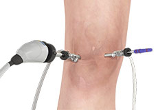
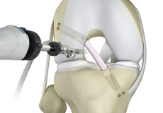
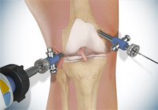
Knee Arthroscopy

Knee arthroscopy is a common surgical procedure performed using an arthroscope, a viewing instrument, to diagnose or treat a knee problem. It is a relatively safe procedure and you will usually be discharged from the hospital on the same day of surgery.
-
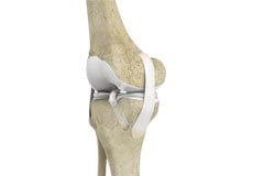
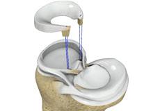
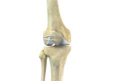
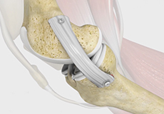
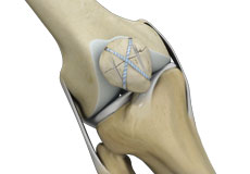
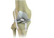
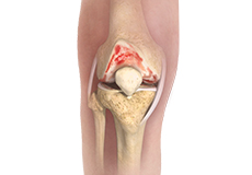
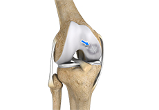
ACL Reconstruction
ACL (anterior cruciate ligament) reconstruction is a commonly performed surgical procedure. With recent advances in arthroscopic surgery, it can now be performed with minimal incision and low complication rates.
-
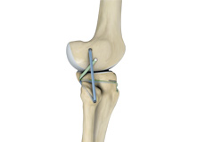
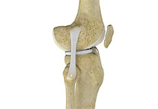
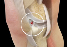
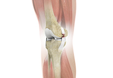
Anterior Cruciate Ligament (ACL) Revision Surgery
The anterior cruciate ligament, or ACL, is one of the major ligaments of the knee that is located in the middle of the knee and runs from the femur (thigh bone) to the tibia (shin bone). When this ligament tears, it does not heal by itself and often leads to the feeling of instability in the knee. Therefore ACL reconstruction surgery becomes necessary. In some people, a second surgery, ACL revision surgery which involves placement of new graft may be needed when ACL reconstruction failure occurs.
-
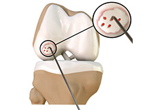
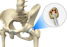
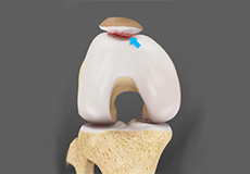
Chondroplasty
Chondroplasty is a surgical procedure to repair and reshape damaged cartilage in a joint. The procedure involves smoothing degenerative cartilage and trimming any unstable flaps of cartilage.
-
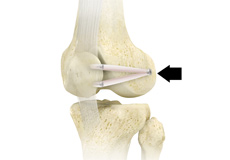
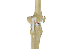
Distal Realignment Procedures
Distal realignment procedures, also known as TTT or tibial tubercle transfer procedures are performed to reposition the kneecap by realigning the tendon under the kneecap to the underlying tibial tubercle. Tibia tubercle is the bony lump on the tibia (bone in the lower leg) below the knee cap. This serves as an attachment point for the patellar ligaments, tendons, and muscles. These procedures are done to prevent patellar subluxation or dislocation.
-
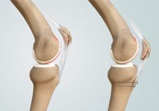
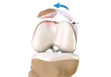
Cartilage Replacement
Cartilage replacement is a surgical procedure performed to replace the worn-out cartilage with new cartilage.
-
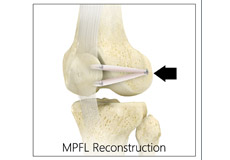
Medial Patellofemoral Ligament Reconstruction
Medial patellofemoral ligament reconstruction is a surgical procedure indicated for severe patellar instability.
-
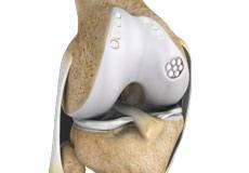
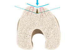
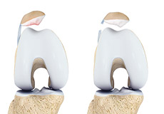
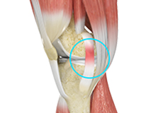
Trochleoplasty
The patella (knee cap) articulates with the lower end of the femur (thigh bone) at the patellofemoral joint. It rests on a groove on the femur called the trochlear groove, which holds it in position while allowing it to glide smoothly during knee movements. Trochlear dysplasia is a condition where the trochlear groove is abnormally shaped, causing the patella to slip out of the groove or dislocate. Trochleoplasty is a surgical procedure that reshapes the trochlea to prevent patellofemoral recurrent instability, and associated pain and disability. The surgery is indicated to treat a trochlea that has a spur (bony outgrowth), is flat or convex.
-
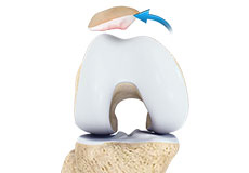
Patellofemoral Knee Replacement
Arthritis is a general term covering numerous conditions where the joint surface or cartilage wears out. The joint surface is covered by a smooth articular surface that allows pain free movement in the joint. This surface can wear out for a number of reasons; often the definite cause is not known.
-
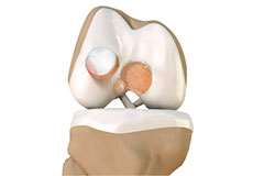
Knee Cartilage Restoration
Knee cartilage restoration is a surgical technique to repair damaged articular cartilage in the knee joint by stimulating new growth of cartilage or by transplanting cartilage into areas with defects in order to relieve pain and restore normal function to the knee.
-
Meniscal Transplantation
Meniscal transplantation is a surgical procedure to replace the damaged meniscus of the knee with healthy cartilage.
-
Joint Realignment
Osteotomy is a surgical procedure wherein a wedge of bone is removed near the degenerated joint and the remaining bone is trimmed and realigned. The surgery aims to ease pain and restore function to the affected joint.
-
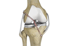
Knee Ligament Reconstruction
Knee ligament reconstruction is a surgical procedure to repair or replace damaged ligaments of the knee joint. The surgery can be performed using minimally invasive techniques.
-
Revision Knee Ligament Reconstruction
Revision knee ligament reconstruction is a complex surgical procedure performed to address failures or to correct the undesirable consequences of primary reconstruction surgery on your knee. The procedure predominantly involves replacing the failed implant or graft with a new one and restoring the stability and functionality of the knee.
-
Posterolateral Corner (PLC) Reconstruction
A posterolateral corner is a complex arrangement of multiple ligaments, tendons, muscles and a joint capsule in your knee. It is located on the outside back corner of the knee.
-
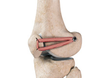
LPFL Reconstruction
LPFL reconstruction or lateral patellofemoral ligament reconstruction is a surgical procedure employed to treat patients with severe patellofemoral instability. The procedure involves replacing a torn lateral patellofemoral ligament with a part of a tendon taken from your leg. The main objective of the LPFL reconstruction is to tighten the knee joint and restore its stability.
-
Multiligament Reconstruction of the Knee
Multiligament knee reconstruction is a surgical procedure to repair or replace two or more damaged ligaments of the knee joint. The surgery can be performed using minimally invasive techniques.
-
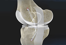
PCL Reconstruction
PCL reconstruction surgery is a procedure to correct torn posterior cruciate ligament (PCL) in the knee using a tissue graft taken from another part of the body, or from a donor.
-
LCL Reconstruction
LCL reconstruction is a surgical procedure to repair torn or damaged lateral collateral ligament in the knee using a tissue graft taken from another part of the body, or from a donor.
-
MCL Reconstruction
MCL reconstruction is a minimally invasive surgical procedure in which a tendon graft is utilized to reconstruct the injured MCL.
-
Knee Trauma Reconstruction
Knee trauma reconstruction is a surgical procedure to repair a soft-tissue injury of the knee such as a torn ligament using a tissue graft or replacing the damaged bony surfaces of the knee joint with an artificial knee joint called a prosthesis. Artificial knee joints are usually made of metal, ceramic or plastic, and consist of the femoral and the tibial components.
-
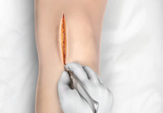
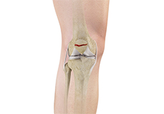
Knee Surgery
Knee surgery is a surgical procedure for the treatment of a knee injury or condition. The procedure involves repairing diseased or damaged structures of the knee joint in order to eliminate pain and restore normal function.
-
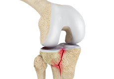
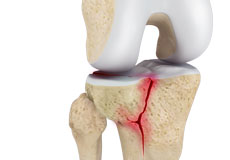
Knee Fracture Surgery
A knee fracture is a broken bone or a crack in or around the joint of the knee. This can involve the tibia (shin bone), the kneecap (patella), or femur (thighbone) where they connect with the knee.
-
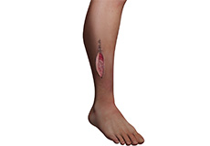
Compartment Decompression
Compartment decompression, also called ‘decompressive fasciotomy’, is a surgical procedure to treat a painful knee condition known as “compartment syndrome”.
-
Patellofemoral Stabilization
Patellofemoral stabilization is a broad term that refers to surgeries employed for stabilization (prevention of dislocation) of the patella for the treatment of patellofemoral instability.
-
Meniscus Replacement
Meniscus replacement is a surgical procedure performed to replace a torn or damaged meniscus in the knee. The meniscus is a soft, fibrous disk of cartilage in your knee joint that sits between the femur (thighbone) and the tibia (shinbone). Also called fibrocartilage, it cushions and stabilizes the joint, acting as a smooth surface for the joint to move on to prevent wear and tear. There are two menisci - one on each side of your knee joint.
-
Patellofemoral Realignment
Patellofemoral realignment is a surgical procedure performed to treat symptomatic patellofemoral instability that does not respond to nonsurgical treatment measures.
-
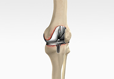
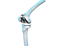
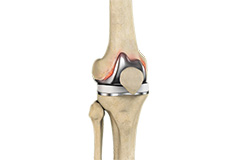
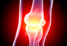
Minimally Invasive Knee Joint Replacement
Total knee replacement is a very successful surgical treatment for knee arthritis. Over the years, minimally invasive knee replacement surgical techniques have been developed to lessen tissue trauma and improve patient outcomes. This minimally invasive approach involves much smaller incisions than the usual 10-12 inch incisions used in the traditional knee replacement and spares the quadriceps muscle and tendon, which control bending of the knee, from being cut to access the knee joint.
-
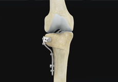
High Tibial Osteotomy
High tibial osteotomy is a surgical procedure performed to relieve pressure on the damaged site of an arthritic knee joint. It is usually performed in arthritic conditions affecting only one side of your knee and the aim is to take pressure off the damaged area and shift it to the other side of your knee with healthy cartilage. During the surgery, your surgeon will remove or add a wedge of bone either below or above the knee joint depending on the site of arthritic damage.
-
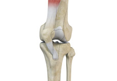
Patellar Tendon Repair
Patellar tendon repair is the surgery performed to reattach the torn tendon to the kneecap and to restore normal function in the affected leg.
-
What is New in Knee Replacement
If you are considering knee replacement surgery, there are new developments under study which can help enhance the quality of life. A suitably-trained surgeon with special skills can offer these procedures.
-
Cartilage Microfracture
Cartilage microfracture is a surgical procedure performed to replace the worn-out articular cartilage with new cartilage
-
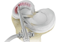
Meniscal Surgery
Meniscal surgery is a surgical procedure employed for the treatment of torn or damaged meniscal tissues in the knee. It is mostly performed as a minimally invasive keyhole procedure.
-
Physeal Sparing Reconstruction of the Anterior Cruciate Ligament
Physeal sparing reconstruction of the anterior cruciate ligament is a surgery to replace a torn anterior cruciate ligament or ACL, a major ligament of the knee, while minimizing damage to the growth plate (physis) present near the end of the bone. Ligaments are powerful bands of tissue that attach one bone to another, and the anterior cruciate ligament attaches the thighbone (femur) to the shinbone (tibia) to help stabilize the knee joint.
-
Cartilage Restoration of the Patellofemoral Joint
The articular or hyaline cartilage is the tissue lining the surface of the two bones in a joint. Cartilage helps the bones move smoothly against each other and can withstand the weight of your body during activities such as running and jumping. Articular cartilage does not have a direct blood supply to it, so has less capacity to repair itself. Once the cartilage is damaged, it will not heal easily and can lead to degeneration of the articular surface, leading to the development of osteoarthritis.
-
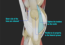
Lateral Lengthening
Lateral lengthening, also known as lateral retinacular lengthening or release, is a surgical procedure to release a tightened lateral retinaculum on the outer aspect of the knee. This procedure is mostly performed to treat knee pain or patellofemoral instability related to chronic pulling of the patella (kneecap) to the outer aspect of the knee, and the inability of the patella to rest properly in the center of the femoral groove as the knee bends and straightens.
-
Physical Therapy for Knee
Physical therapy is an exercise program that helps you to improve movement, relieve pain, encourage blood flow for faster healing, and restore your physical function and fitness level. It can be prescribed as an individual treatment program or combined with other treatments. It involves a combination of education, manual therapy, exercises and techniques such as water, heat, cold, electrical stimulation and ultrasound.
-
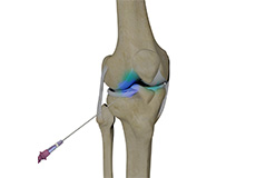
Intraarticular Knee Injection
Knee pain and stiffness can be disabling and difficult to treat. It can limit an individual’s lifestyle and negatively impact body image and emotional well-being.
-
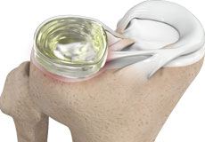
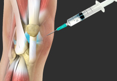
Viscosupplementation
Viscosupplementation refers to the injection of a hyaluronan preparation into the joint. Hyaluronan is a natural substance present in the joint fluid that assists in lubrication. It allows the smooth movement of the cartilage-covered articulating surfaces of the joint.


-
Knee Arthritis
The joint surface is covered by a smooth articular surface that allows pain-free movement in the joint. Arthritis is a general term covering numerous conditions where the joint surface or cartilage wears out. This surface can wear out for several reasons; often the definite cause is not known. Arthritis often affects the knee joint. When the articular cartilage wears out, the bone ends rub on one another and cause pain. The most common type of arthritis is osteoarthritis. It occurs with aging and use.
-
Knee Osteoarthritis
Osteoarthritis also called degenerative joint disease, is the most common form of arthritis. It occurs most often in older people. This disease affects the tissue covering the ends of bones in a joint (cartilage).In a person with osteoarthritis, the cartilage becomes damaged and worn out causing pain, swelling, stiffness and restricted movement in the affected joint. This condition most commonly affects the joints in the hips, knees, hands, and spine. Rarely, the disease may affect the shoulders, wrists, and feet.
-
Patellofemoral Arthritis
Patellofemoral arthritis is an inflammatory condition characterized by loss of the smooth cartilage between the kneecap (patella) and the underlying femoral (thigh) bone in the knee joint. When the articular cartilage wears out, the underlying bones rub against each other, causing pain, swelling, stiffness, and restricted movement.
-
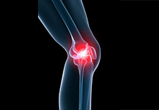
Knee Pain
Knee pain is a common condition affecting individuals of various age groups. It not only affects movement but also impacts your quality of life. An injury or disease of the knee joint or any structure surrounding the knee can result in knee pain. A precise diagnosis of the underlying cause is important to develop an appropriate treatment plan.
-
Knee Fracture
A fracture is a condition in which there is a break in the continuity of the bone. In younger individuals, these fractures are caused by high energy injuries, as from a motor vehicle accident. In older people, the most common cause is a weak and fragile bone.
-
Fractures of the Patella
The patella or kneecap is a small bone present in the front of your knee where the thigh bone meets the shinbone. It provides protection to your knee and attachment to muscles in the front of the thigh. An injury to the knee can result in a break or fracture of the patella.
-
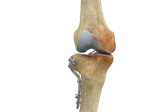
Periprosthetic Knee Fractures
Knee replacement, also called knee arthroplasty, is a surgical procedure in which the worn-out or damaged surfaces of the knee joint are removed and replaced with artificial implants. Any resulting fractures or breaks in the bone around the implant are called periprosthetic knee fractures. These fractures may occur during surgery (intraoperative) or after surgery (postoperative), and usually involve the patella, tibia or the femur (kneecap, shinbone, and thighbone). Women are at higher risk than men.
-
Fractures of the Tibia
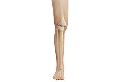
The lower leg is made up of two long bones called the tibia and fibula that extend between the knee and ankle. The tibia or shinbone is the larger of the two bones.
-
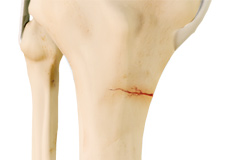
Tibial Plateau Fracture
A tibial plateau fracture is a crack or break on the top surface of the tibia or shinbone in the knee joint. The fracture most often occurs following a high-intensity trauma or injury from the impaction of the femoral condyles over the tibial plateau. Fractures of the tibial plateau are serious injuries that are more commonly seen in athletes involved in high-impact sports such as basketball, rugby, and football and can put athletes out of action for a long period of time.
-
Tibial Eminence Fracture
The tibia or shin bone is a major bone of the leg which connects the knee to the ankle. A fracture or break in the upper part of the tibia is known as a proximal tibial fracture and commonly occurs just below the knee joint.
-
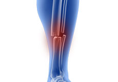
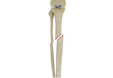
Tibial Shaft Fracture
A tibial shaft fracture is a crack or break in the middle section of the tibia bone due to severe trauma. The lower leg is made up of two long bones called the tibia and fibula that extend between the knee and ankle and help form the ankle joint and knee joint. The tibia or shinbone is the larger of the two bones and is one of the major bones of the lower leg. It bears most of the body’s weight and plays a crucial role in balancing body weight when standing and walking. The tibia is the most frequently fractured long bone of the body. It normally takes a great amount of force for a fracture of the tibia to occur.
-
Knee Stress Fractures
Stress fractures of the patella or knee are very rare. Approximately two out of 10,000 athletes may experience a patella stress fracture. Initial symptoms include activity-related pain and then a fatigue stress fracture after minor trauma. The term insufficiency stress fracture is used for cases where the patella is weakened previously such as after patella resurfacing surgery.
-
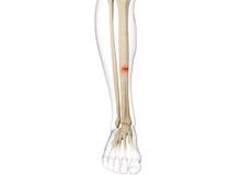
Stress Fracture of the Tibia
A stress fracture of the tibia or shinbone is a thin fracture, also called a hairline fracture that occurs in the tibia due to excess stress or overuse. The tibia is a weight-bearing bone in which stresses can accumulate from activities such as running and jumping.
-
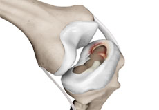
Meniscal Tears
A meniscal tear is a common knee injury in athletes, especially those involved in contact sports. A sudden bend or twist in your knee causes the meniscus to tear. Elderly people are more prone to degenerative meniscal tears as the cartilage wears out and weakens with age.
-
ACL Tears
The anterior cruciate ligament (ACL) is one of the major ligaments of the knee. It is located in the middle of the knee and runs from the femur (thighbone) to the tibia (shinbone). The ACL prevents the tibia from sliding out in front of the femur. Together with the posterior cruciate ligament (PCL), it provides rotational stability to the knee.
-
Knee Injury
Pain, swelling, and stiffness are the common symptoms of any damage or injury to the knee. If care is not taken during the initial phases of injury, it may lead to joint damage, which may end up destroying your knee.
-
Knee Ligament Injuries
Knee problems may arise if any of these structures get injured by overuse or suddenly during sports activities. Pain, swelling, and stiffness are the common symptoms of any damage or injury to the knee. The common causes of knee injury include:
-
Meniscal Injuries
Meniscal tears are one of the most common injuries to the knee joint. It can occur at any age but are more common in athletes involved in contact sports. The meniscus has no direct blood supply and for that reason, when there is an injury to the meniscus, healing is difficult.
-
Articular Cartilage Injury
Articular or hyaline cartilage is the tissue lining the surface of the two bones in the knee joint. Cartilage helps the bones move smoothly against each other and can withstand the weight of the body during activities such as running and jumping. Articular cartilage does not have a direct blood supply to it so has little capacity to repair itself. Once the cartilage is torn it will not heal easily and can lead to degeneration of the articular surface, leading to the development of osteoarthritis.
-
Terrible Triad Injuries
Terrible triad injury, also known as Unhappy triad or O’Donoghue triad, is a condition involving injury to three structures in the knee joint.
-
Knee Sports Injuries
Trauma is any injury caused during physical activity, motor vehicle accidents, electric shock, or other activities. Sports trauma or sports injuries refer to injuries caused while playing indoor or outdoor sports and exercising. Sports trauma can result from accidents, inadequate training, improper use of protective devices, or insufficient stretching or warm-up exercises. The most common sports injuries are sprains and strains, fractures, and dislocations.
-
Multiligament Knee Injuries
Injury to more than one knee ligament is called a multiligament knee injury and may occur during sports or other physical activities.
-
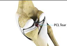
PCL Injuries
Posterior cruciate ligament (PCL), one of the four major ligaments of the knee, is situated at the back of the knee. It connects the thighbone (femur) to the shinbone (tibia). The PCL limits the backward motion of the shinbone. PCL injuries are very rare and more difficult to detect than other knee ligament injuries. Cartilage injuries, bone bruises, and ligament injuries often occur in combination with PCL injuries.
-
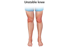
Unstable Knee
Damage to any of these supportive structures causes instability of the knee joint. An unstable knee can be caused by the sudden twisting of the knee, tears of the meniscus, ligament or capsule, osteoarthritis of the knee (wear and tear of the cushioning cartilage tissue between the bones) and sports injuries. When these tissues get injured, the patella or kneecap can move out of its groove in the knee joint and lead to instability.
-
Chondromalacia Patella
Chondromalacia patella is a common condition characterized by softening, weakening and damage of the cartilage. The condition is most often seen in young athletes and older adults who have arthritis of the knee. It especially occurs in women.
-
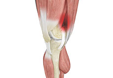
Jumper's Knee
Jumper’s knee, also known as patellar tendinitis, is inflammation of the patellar tendon that connects your kneecap (patella) to your shinbone. This tendon helps in the extension of the lower leg.
-
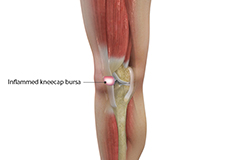
Kneecap Bursitis
Bursitis refers to the inflammation and swelling of the bursa. Inflammation of the bursa in front of the kneecap (patella) is known as kneecap bursitis or prepatellar bursitis.
-
Iliotibial Band Syndrome
Iliotibial band syndrome is an overuse injury resulting from the inflammation of the iliotibial band. It occurs when the iliotibial band and the lower outside portion of the thighbone at the knee joint rub against each other.
-
Osteochondritis Dissecans of the Knee
Osteochondritis dissecans is a joint condition in which a piece of cartilage, along with a thin layer of the bone separates from the end of the bone because of inadequate blood supply. The separated fragments are sometimes called “joint mice”. These fragments may be localized or may detach and fall into the joint space, causing pain and joint instability. Osteochondritis dissecans occurs within the lateral aspect of the medial femoral condyle. The condition can also occur in other joints, including your elbows, ankles, shoulders, and hips.
-
Medial Patellofemoral Ligament (MPFL) Tears
The medial patellofemoral ligament (MPFL) is a ligament that joins the kneecap (patella) to the thighbone (femur). Ligaments are fibrous connective tissue that attach bones to other bones. The MPFLis attached to the inside of the patella and helps stabilize it and position it in a groove at the lower end of the femur called the trochlea. Trauma or a blow to the knee may cause a tear in the ligament.
-
Knee Sprain
Knee sprain is a common injury that occurs from overstretching of the ligaments that support the knee joint. A knee sprain occurs when the knee ligaments are twisted or turned beyond its normal range, causing the ligaments to tear.
-
MCL Tears
The medial collateral ligament (MCL) is the ligament located on the inner part of the knee joint. It runs from the femur (thighbone) to the top of the tibia (shinbone) and helps in stabilizing the knee.
-
MCL Sprains
MCL sprains occur due to a sudden impact from the outside of your knee, most commonly while playing sports such as rugby and football. Rarely, the MCL can get injured when the knee gets twisted or following a quick change in direction.
-
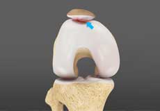
Patellar Dislocation/Patellofemoral Dislocation
Patellar dislocation occurs when the patella moves out of the patellofemoral groove, (trochlea) onto the bony head of the femur. If the kneecap partially comes out of the groove, it is called subluxation; if the kneecap completely comes out, it is called dislocation (luxation).
-
Recurrent Patella Dislocation
The patella (kneecap) is a small bone that shields your knee joint. It is present in front of your knee, on a groove called the trochlear groove that sits at the junction of the femur (thighbone) and tibia (shinbone). Articular cartilage presents below the patella and end of the femur cushion and helps the bones glide smoothly over each other when the legs move. This joint is stabilized and supported by a network of soft tissues. The medial patellofemoral ligament (MPFL) connects to the inner side of the patella and helps to keep it from slipping away from the knee. Damage to this ligament leads to patellar dislocation.
-
Quadriceps Tendon Rupture
The quadriceps can rupture after a fall, direct blow to the leg and when you land on your leg awkwardly from a jump. Quadriceps tendon rupture most commonly occurs in middle-aged people who participate in sports that involve jumping and running. Other causes include tendonitis (inflammation of quadriceps tendon), diseases such as rheumatoid arthritis, diabetes mellitus, infection and chronic renal failure, which weaken the quadriceps tendon. Use of medications such as steroids and some antibiotics also weakens the quadriceps tendon.
-
Patellar Tendon Rupture
The patellar tendon works together with the quadriceps muscle and the quadriceps tendon to allow your knee to straighten out. Patella tendon rupture is the rupture of the tendon that connects the patella (kneecap) to the top portion of the tibia (shinbone).
-
Lateral Meniscus Syndrome
Lateral meniscus syndrome is characterized by an injury caused by the tearing of the cartilage tissue or a rare case of a congenital abnormality called a discoid meniscus, which results in knee pain.
-
Patellar Instability
Any damage to the supporting ligaments may cause the patella to slip out of the groove either partially (subluxation) or completely (dislocation). This misalignment can damage the underlying soft structures such as muscles and ligaments that hold the kneecap in place. Once damaged, these soft structures are unable to keep the patella (kneecap) in position. Repeated subluxation or dislocation makes the knee unstable. This condition is called knee instability. Patellar (kneecap) instability results from one or more complete or partial dislocations (subluxations).
-
Multiligament Instability
The knee is a complex joint of the body that is vital for movement. The four major ligaments of the knee are anterior cruciate ligament (ACL), posterior cruciate ligament (PCL), medial collateral ligament (MCL) and lateral collateral ligament (LCL). They play an important role in maintaining the stability of the knee. A multiligament injury is a tear in one or more ligaments of the knee, which affects the knee stability.
-
Patellofemoral Instability
Patellofemoral instability means that the patella (kneecap) moves out of its normal pattern of alignment. This malalignment can damage the underlying soft structures such as muscles and ligaments that hold the knee in place.
-
Posterolateral Instability
Posterolateral instability, also known as posterolateral rotatory instability (PLRI), is a common pattern of knee instability that results from injuries to the structures that support the outside of the knee joint, the posterolateral corner. When individuals injure the knee ligaments, they are most likely to injure the structures of the posterolateral corner. These structures include the popliteus tendon, lateral collateral ligament, and the knee joint capsule. The knee instability mostly occurs along with other ligamentous injuries, in particular, damage to either the ACL (anterior cruciate ligament), the PCL (posterior cruciate ligament), or both.
-
Lateral Patellar Instability
Lateral patellar instability is defined as a lateral shift or displacement of the patella (kneecap) as a result of disruptive changes in the medial patellofemoral ligament (MPFL) and medial patellar retinaculum.
-
Medial Patellar Instability
Medial patellar instability is a disabling condition characterized by medial subluxation of the patella which occurs as a complication of lateral retinacular release surgery. Lateral retinacular release is a surgical procedure employed for the treatment of lateral patellar instability. However, disruption of the lateral patellar structures during this surgery has been associated with medial patellar instability.
-
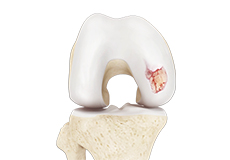
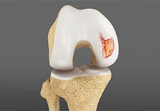
Osteochondral Defect of the Knee
An osteochondral defect, also commonly known as osteochondritis dissecans, of the knee refers to a damage or injury to the smooth articular cartilage surrounding the knee joint and the bone underneath the cartilage. The degree of damage may range from a rupture of the cartilage to a slight crack of the bone to a piece of the bone breaking off within the joint. These pieces may vary in size and depth and may remain stable or unstable within the joint. These may occur from an acute traumatic injury to the knee or an underlying disorder of the bone. Osteochondral defect is more common among young athletes who actively take part in sports and most commonly affects the femoral condyles in the knee.
-
Knee Dislocation
Knee dislocation is a condition that occurs when the bones that form the knee joint, namely the femur or thigh bone get separated from the shin bone. This can cause serious damage to the nerves, blood vessels, and ligaments surrounding the knee, leading to a decline in strength and overall health of the leg.
-
Chronic Hamstring Tendinopathy
Chronic hamstring tendinopathy is a condition characterized by a deep pain in the buttocks and upper part of the back of the thigh. It is also referred to as chronic high hamstring tendinopathy or proximal hamstring tendinopathy.
-
Patellar Tracking Disorder/Patellar Maltracking
Patellar tracking disorder, also known as patellar maltracking, is a condition in which the kneecap (patella) moves sideways from its groove when the leg is bent or straightened.
-
Patellar Tendinitis
Patellar tendinitis, also known as "jumper's knee", is an inflammation of the patellar tendon that connects your kneecap (patella) to your shinbone. This tendon helps in extension of the lower leg.
-
Meniscus Root Tear
Meniscal root tears are characterized as soft tissue or bony root avulsion injuries or radial tears located within 1 cm of meniscus root attachment. They can be either a tear which disconnects the root area completely from the body of the meniscus (complete radial tear) or a disruption of the meniscus attachment directly from the bone (true meniscus root tear) that can cause the whole meniscus to lose its capacity to safeguard the underlying cartilage.
-
Women and ACL Injuries
The anterior cruciate ligament is one of the four major ligaments of the knee that connects the femur (thigh bone) to the tibia (shin bone) and helps stabilize the knee joint. Anterior cruciate ligament (ACL) injury is one of the common injuries of the knee. An injury to the ACL commonly occurs during sports or activities that involve twisting, overextension, landing from a jump incorrectly and abrupt change in direction or speed of movements.
-
Quadriceps Tendon Rupture and Repair
A quadriceps tendon rupture is defined as a tear of the quadriceps tendon as a result of a traumatic incident. The quadriceps tendon is a strong rope-like fibrous tissue located at the top of the patella or kneecap that connects the quadriceps muscles to the kneecap. It works together with the quadriceps muscles to allow us to straighten our leg. The quadriceps muscles are the muscles located at the front of the thigh.
-
Anterior Knee Pain
Anterior knee pain is characterized by chronic pain over the front and center of the knee joint. It is common in athletes, active adolescents (especially girls) and overweight individuals. Anterior knee pain refers to various conditions, which include runner's knee or patellar tendinitis, and chondromalacia of the patella. There is an inter-individual variation in the duration and presentation of pain.
-
Osgood-Schlatter Disease
Osgood-Schlatter disease refers to a condition in older children and teenagers caused by excessive stress to the patellar tendon (located below the kneecap). Participants in sports such as soccer, gymnastics, basketball, and distance running are at higher risk for this disease.
The knee is a complex joint made up of different structures - bones, tendons, ligaments, and muscles. They all work together to maintain the knee’s normal function and provide stability to the knee during movement.
Having a well-functioning healthy knee is essential for our mobility and ability to participate in various activities. Understanding the anatomy of the knee enhances your ability to discuss and choose the right treatment procedure for knee problems with your doctor.
Bones of the Knee
The knee is a hinge joint made up of two bones, the thighbone (femur) and shinbone (tibia). There are two round knobs at the end of the femur called femoral condyles that articulate with the flat surface of the tibia called the tibial plateau. The tibial plateau on the inside of the leg is called the medial tibial plateau and on the outside of the leg, the lateral tibial plateau.
The two femoral condyles form a groove on the front (anterior) side of the knee called the patellofemoral groove. A small bone called the patella sits in this groove and forms the kneecap. It acts as a shield and protects the knee joint from direct trauma.
A fourth bone called the fibula is the other bone of the lower leg. This forms a small joint with the tibia. This joint has very little movement and is not considered a part of the main joint of the knee.
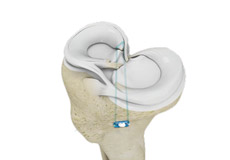
Articular Cartilage and Menisci of the Knee
Movement of the bones causes friction between the articulating surfaces. To reduce this friction, all articulating surfaces involved in the movement are covered with a white, shiny, slippery layer called articular cartilage. The articulating surface of the femoral condyles, tibial plateaus and the back of the patella are covered with this cartilage. The cartilage provides a smooth surface that facilitates easy movement.
To further reduce friction between the articulating surfaces of the bones, the knee joint is lined by a synovial membrane that produces a thick clear fluid called synovial fluid. This fluid lubricates and nourishes the cartilage and bones inside the joint capsule.
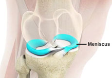
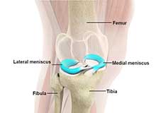
Within the knee joint, between the femur and tibia, are two C-shaped cartilaginous structures called menisci. Menisci function to provide stability to the knee by spreading the weight of the upper body across the whole surface of the tibial plateau. The menisci help in load-bearing i.e. it prevents the weight from concentrating onto a small area, which could damage the articular cartilage. The menisci also act as a cushion between the femur and tibia by absorbing the shock produced by activities such as walking, running and jumping.
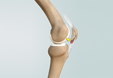
Ligaments of the Knee
Ligaments are tough bands of tissue that connect one bone to another bone. The ligaments of the knee stabilize the knee joint. There are two important groups of ligaments that hold the bones of the knee joint together, collateral and cruciate ligaments.
Collateral ligaments are present on either side of the knee. They prevent the knee from moving too far during side to side motion. The collateral ligament on the inside is called the medial collateral ligament (MCL) and the collateral ligament on the outside is called the lateral collateral ligament (LCL).
Cruciate ligaments, present inside the knee joint, control the back-and-forth motion of the knee. The cruciate ligament in the front of the knee is called anterior cruciate ligament (ACL) and the cruciate ligament in the back of the knee is called posterior cruciate ligament (PCL).
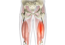
Muscles of the Knee
There are two major muscles in the knee - the quadriceps and the hamstrings, which enable movement of the knee joint. The quadriceps muscles are located in front of the thigh. When the quadriceps muscles contract, the knee straightens. The hamstrings are located at the back of the thigh. When the hamstring muscles contract, the knee bends.
Tendons of the Knee
A tendon is a tissue that attaches a muscle to a bone. The quadriceps muscles of the knee meet just above the patella and attach to it through a tendon called the quadriceps tendon. The patella further attaches to the tibia through a tendon called the patella tendon. The quadriceps muscle, quadriceps tendon, and patellar tendon all work together to straighten the knee. Similarly, the hamstring muscles at the back of the leg are attached to the knee joint with the hamstring tendon.

