Adam B. Yanke, MD is a sports medicine orthopedic surgeon with a special interest in advanced arthroscopy, shoulder replacement, and a special focus on patellofemoral dysfunction and cartilage restoration. Click on the below tabs to know more about his services.
Sports Medicine
Sports Medicine, also known as sports and exercise medicine (SEM), is a branch of medicine that deals with the treatment and prevention of sports and exercise-related injuries and improving fitness and performance.
Chicago Riot Rugby
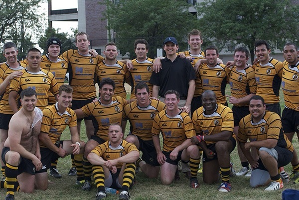
Head Team Physician
Affiliated Physician
 Chicago Bulls
If you wish to be advised on the most appropriate treatment, please call to schedule an appointment or click to request an appointment online.

